
How a cloud experiment in Uganda could redefine global health
The collaboration between Institute of Instrumentation for Molecular Imaging (I3M) in Spain and Mbarara University of Science and Technology in Uganda began with shared ambition and a shared limitation. The teams were working together on applied MRI research for Sub-Saharan Africa, but the images they could produce were not always reliable enough to interpret.
The MRI system in use could capture signals, but turning those signals into clear images was another matter. Noise crept in, detail washed out. The processing required to make sense of the data exceeded what the local system could handle, slowing experimentation and limiting what the team could reliably see.
Looking for a way forward, the researchers adopted Tyger (opens in new tab), an open-source platform developed by Microsoft Research. With Tyger moving the most computationally intensive stages off the scanner and into the cloud, and denoising models such as SNRAware—also developed by Microsoft Research—refining the output, the team was able to produce images that reached a level of clarity suitable for interpretation.
Because the heavy computation happens remotely with Tyger, teams can test, iterate, and develop new imaging approaches without being constrained by the capabilities of local hardware. That means they can try ideas, see results, and adjust course in hours rather than days or weeks, without waiting on machines to catch up. Tyger is now being evaluated across a range of MRI systems in research environments where that flexibility matters.
Where reconstruction does the work
For Michael Hansen, General Manager of Medical Imaging at Microsoft Research Health Futures, and his team, Tyger was born from a deep curiosity about what medical instruments might become when their limitations are no longer dictated by the hardware inside them.
Hansen often describes conventional MRI machines as creatures shaped by their magnets: massive, power-hungry, and dependent on the thick walls and high current of large hospitals. The physics of the magnet has always set limitations for images. But the team began to see a way to unsettle that hierarchy.
“If you have lots of compute, you can make compromises on the instrument side. You can change one currency for another.”
– Michael Hansen, Microsoft Health Futures
In other words, if reconstruction is no longer tied to a specific class of hardware, software and computation can carry work that magnets and electronics once had to bear.
Tyger formalizes that tradeoff across MRI systems operating at different field strengths. It allows scanners, whether high field, low field, or ultra-low field, to function primarily as signal gathering devices, with reconstruction handled in the cloud. The lineage of MRI—in which image quality has historically been secured through greater size and cost—is inverted. Less infrastructure, more intelligence.
In the i3M–Mbarara collaboration, this approach is applied in an ultra-low field setting, without redesigning or replacing the MRI device itself. Researchers work with the scanner available to them and focus instead on how its data can be reconstructed more effectively.
The raw electrical signals from the scanner travel, sometimes over a barely adequate mobile network, to Azure, Microsoft’s trusted cloud platform. There, denoising models like SNRAware are put to work. Distortion correction algorithms draw on prior knowledge of how a low magnetic field bends and warps the signal. The computational load that would overwhelm a compact industrial PC at the scanner is absorbed by the Tyger environment, which provides a depth of compute that once required entire rooms of hardware.
What comes back is an image shaped by reconstruction and computation, rather than the physical limits of the scanner that acquired it.
A real-world test of interpretability
At Mbarara University of Science and Technology, the limitations of MRI reconstruction will shape what clinicians can diagnose and how long patients will wait for answers.
In Uganda, where injury and illness routinely outstrip the reach of advanced medical equipment, the distance between emergency and diagnosis can stretch unbearably long.
Many patients must travel long distances to reach facilities with imaging equipment, and in some cases the nearest MRI scanner is hundreds of kilometers away. When images are unclear or need to be repeated, the cost is measured in time, financial burden, and delayed care.
In this setting, where much of the population has no access to MRI at all, image quality is more than a matter of optimization. The consequences cascade through everyday life: stroke patients lose precious hours without diagnosis, children with hydrocephalus, a dangerous buildup of fluid in the brain, are identified only when their heads have already swollen. Trauma victims are treated by experience rather than tangible evidence.
“For many patients who cannot afford or manage travel to distant hospitals, the situation is devastating. We have only one MRI imaging facility outside the Capital City, lower-level facilities have neither MRI nor CT imaging. Technology like this [Tyger] could finally bring imaging to the places where it has never existed.”
— Eng. Dr. Johnes Obungoloch, Dean of the Faculty of Applied Sciences and Technology, Mbarara University of Science and Technology
From Arizona to Uganda
The inspiration to use Tyger for ultra-low-field MRI took shape in 2023 at a reconstruction workshop in Arizona, where a conversation about the challenges of MRI in low resource settings left Hansen thinking about the limits of expertise. He recalls thinking that methods honed for pushing the limits of high-end scanners don’t always translate cleanly to environments with tighter constraints—and that rethinking reconstruction there can call for a different set of assumptions and tradeoffs. Hansen’s realization was that the problem statement itself had to change, and with it, who might be best equipped to solve it.
After the session, Spanish physicist Joseba Alonso approached him. Alonso, a researcher at the i3M institute in Valencia, had been thinking about what MRI might look like beyond the controlled conditions of major research hospitals. Soon enough, his team started preparing to travel to Mbarara University of Science and Technology, where colleagues were working with an MRI system under far tighter constraints.
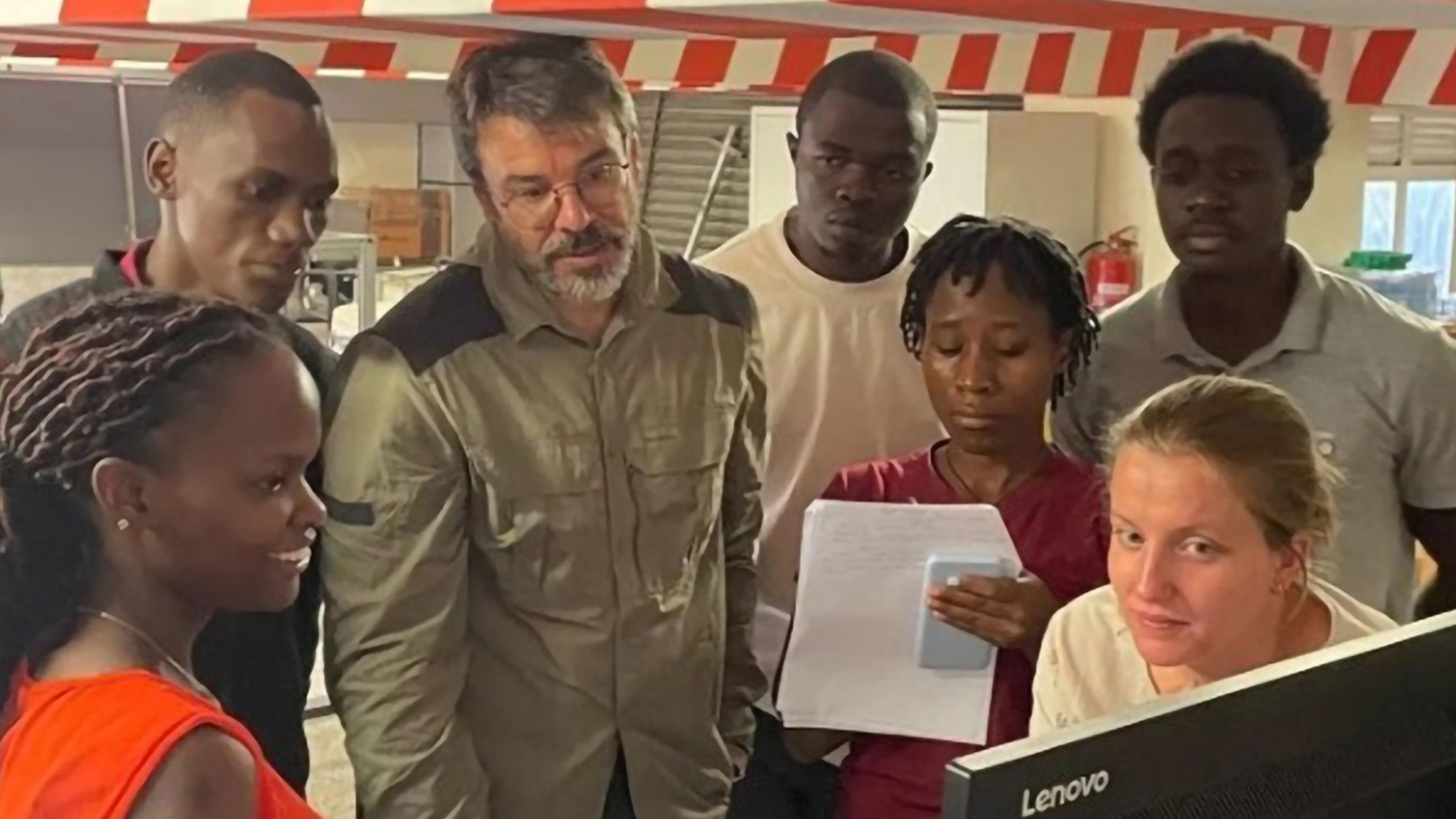
The connection proved consequential. What began as a conversation turned into a set of practical experiments. Researchers at i3M and Mbarara started exchanging data, testing reconstruction approaches, and linking the scanner in Uganda to Tyger’s cloud-based pipeline. The early work was informal and provisional, shaped by distance and limited infrastructure, but it held. Today, i3M and Mbarara University collaborate closely, using Tyger as a shared foundation for their MRI research.
“There is synergy between our goals,” Alonso said. “We want to produce locally the knowledge to build and use these machines.”
At Mbarara University, the work has also become an educational effort. Dr. Obungoloch and his colleagues train engineering and medical students, as well as community healthcare workers. Many trainees who had never seen an MRI before are now working to make improvements on the low-field machines in use.
“We have scanned dozens of volunteers since summer 2025,” he said. “At first, we could only get part of the head. Now we can acquire full head images.”
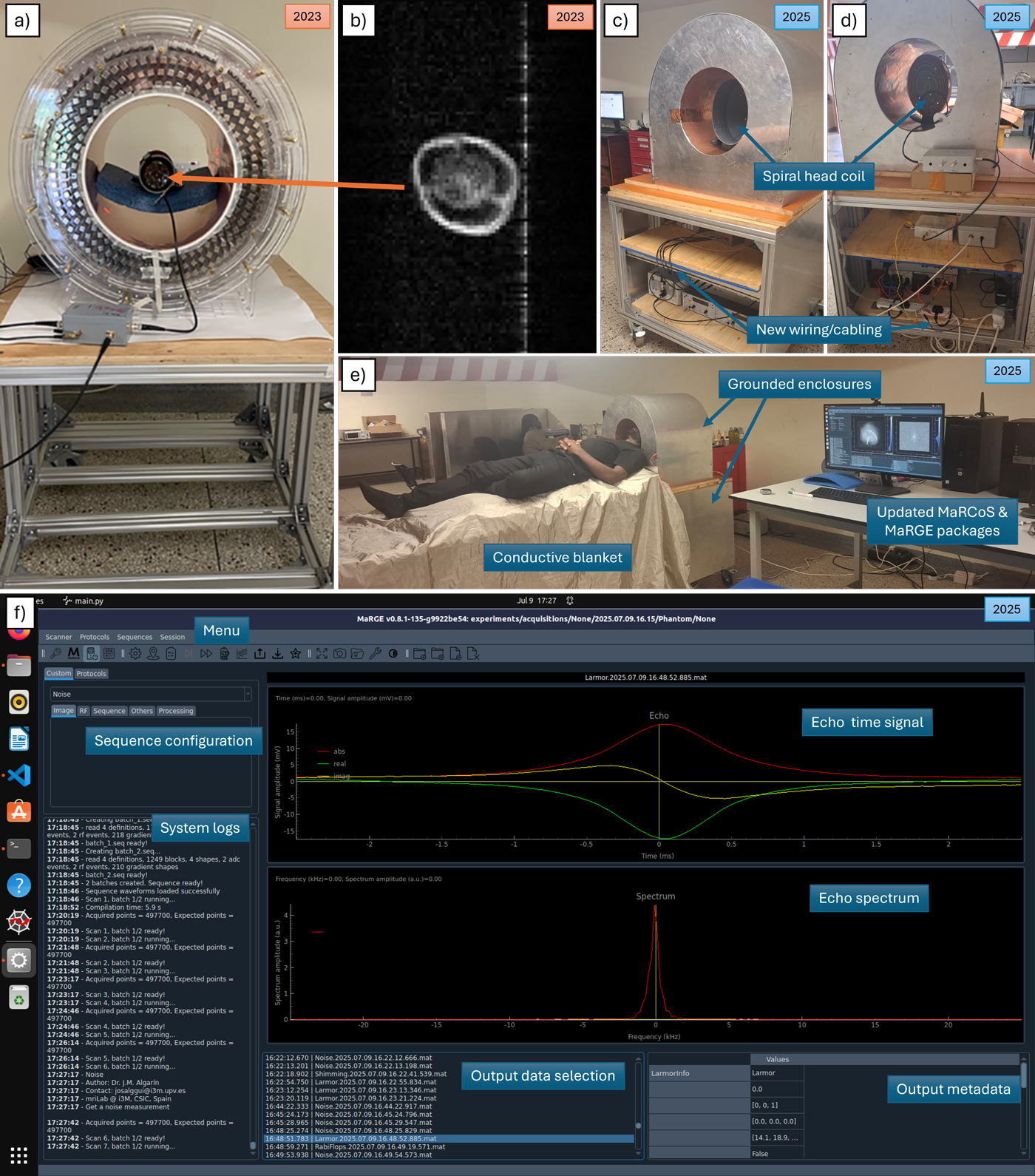
b) The first reconstructed image, a bell pepper, which took several hours to process.
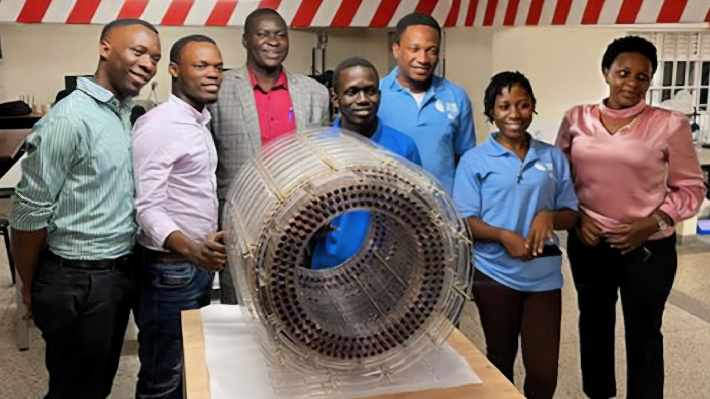
c–e) Front, rear, and full views of the scanner in its current configuration, showing improvements to the electronics, RF coils, and mechanical integration.
f) The MaRGE user interface used to operate the system.
Some students are already moving on to advanced degrees abroad. Others are gaining skills in electronics, design, and signal processing—competencies that extend far beyond MRI.
What the workflow makes possible
Hansen offered an analogy that neatly captures the moment Tyger occupies: Early cellphone cameras were first seen as limited and unserious, yet they evolved into the most widely used imaging devices in the world. Tools that begin modestly can, under the right conditions, redefine a field.
Realizing that potential will require more than technical progress. As Alonso noted, the transition to clinical systems depends on regulatory approval, manufacturing capacity, and the ability to support devices locally.
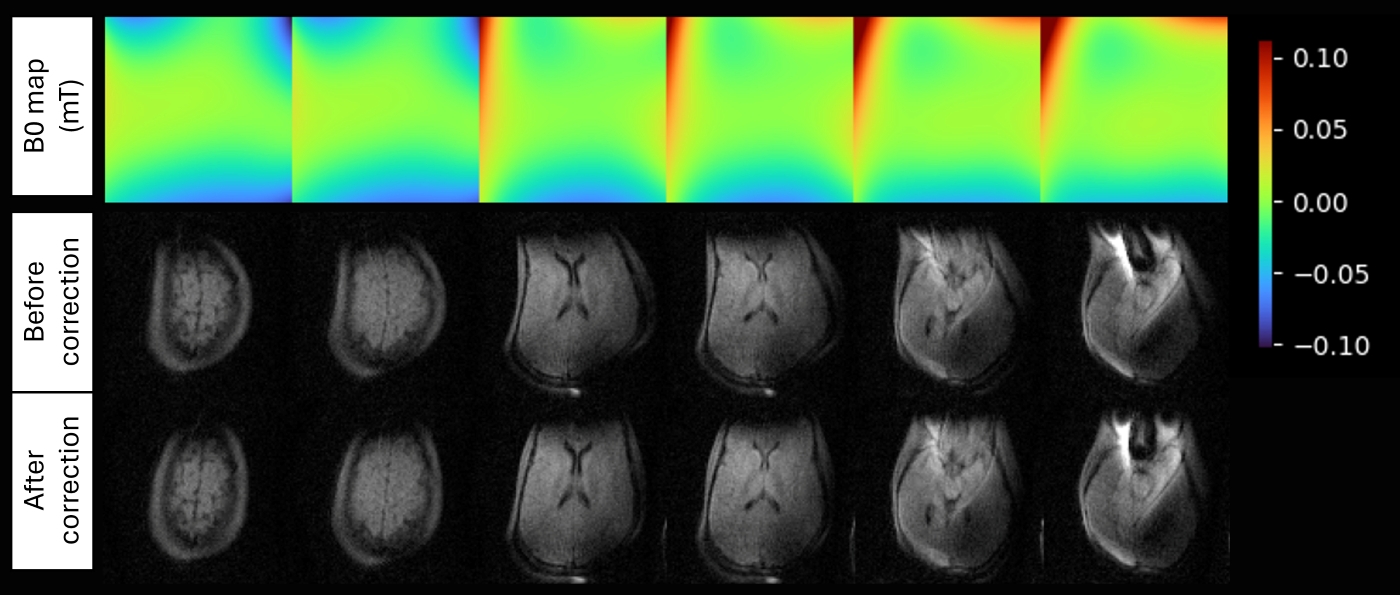
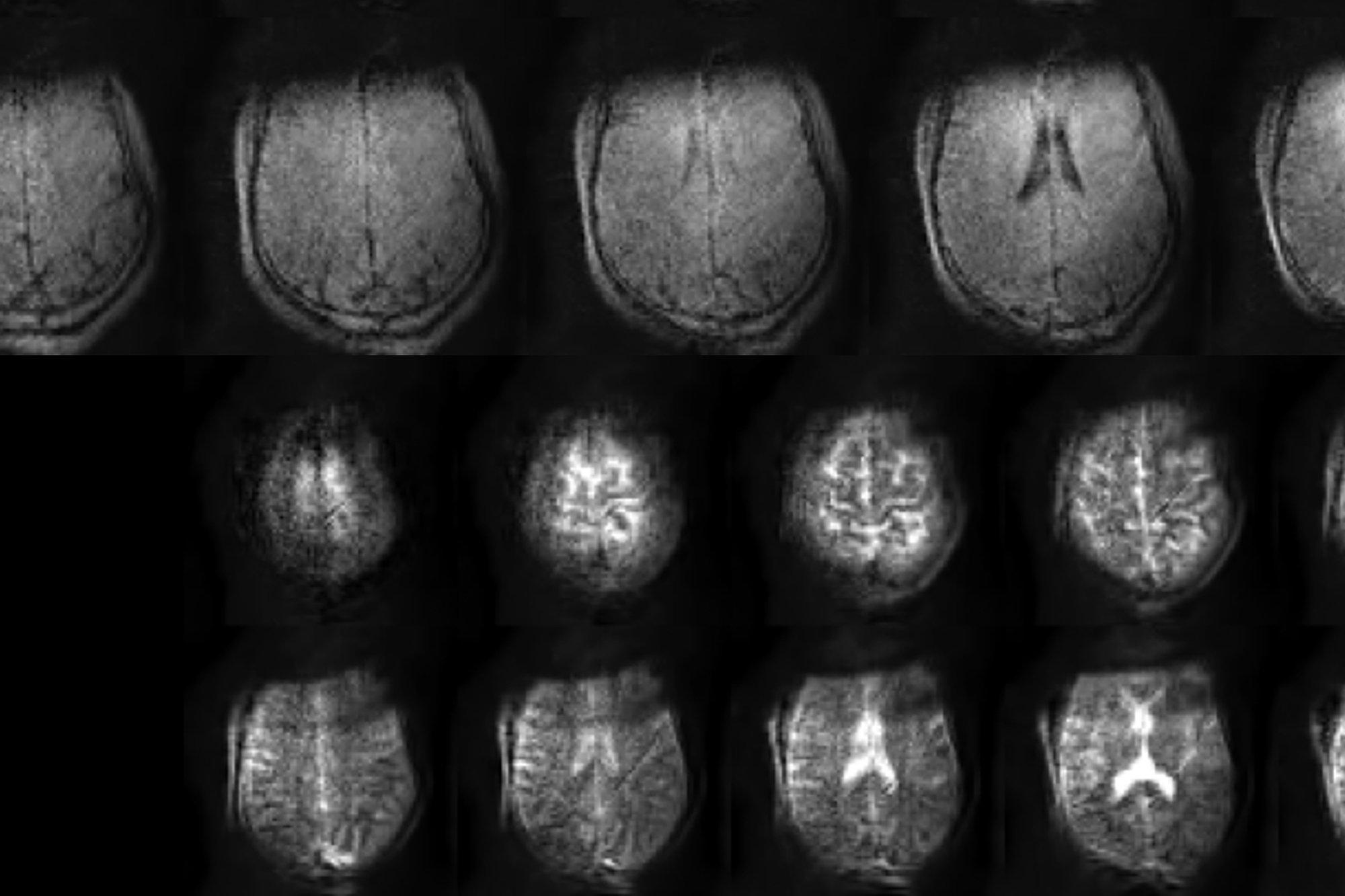
Middle row: A conventional image reconstruction that does not account for these field variations, resulting in visible geometric distortion.
Bottom row: A Tyger reconstruction that incorporates the measured field map, producing a clearer image with substantially reduced distortion, especially in the frontal regions of the brain.
Still, Tyger changes what is technically feasible. By shifting computational complexity to the cloud, it creates an architecture in which hardware, software, and imaging workflows reinforce rather than compete with one another. Hansen described it as “the connective tissue that enables practical impact,” because it allows imaging systems to rely on shared computation rather than on local hardware alone.
The collaboration between i3M and Mbarara offers a concrete example of how that plays out in practice. The field is shifting. Increasingly, image quality depends less on where a scan is performed and more on how its data is reconstructed. Software is key to making this happen.
Advancing AI to meet needs of the global majority
Generative AI powers apps and tools that boost productivity and knowledge in much of the world.
But these systems don’t work equally well for all communities—especially those under-represented online, where most AI training data originates. As a result, generative AI performs poorly in many languages and does not reflect the social and cultural realities of every population.
Paza Speech Playbook
A practical guide for building Automatic Speech Recognition (ASR) models that work in the real world.
Atlas Playbook
A practical, grounded guidance for designing, deploying, and evaluating AI across diverse cultural contexts.
Vibhasha Playbook
A comprehensive guide for building language model applications in multilingual and multicultural settings.
3D Telecommunications goes open source
Bringing Holoportation™ from the lab to the field.
Story contributors: Amanda Black, David Celis Garcia, Alyssa Hughes, Lindsay Kalter, Brenda Potts, Lindsay Shanahan, Amber Tingle, Shauna Whooley, Asa-Mari Zephirin